How do Ultrasound scans work?

Ultrasound is a phrase that is almost widely used colloquially, and it is very common among pregnant women. For many years, it has been a very prominent medical imaging technique.
What does the term “Ultrasound” mean?
Ultrasound, or sonography, is a non-invasive technique for collecting images of the inside of the body, including blood vessels, muscles, organs, and other soft tissues. An ultrasonic scan uses high-frequency sound waves to generate images of the interior of the body. During pregnancy it allow the sonographer to examine how the baby is developing within the mother’s womb and to detect any abnormalities.
This scan may be use to evaluate fetal growth and detect abnormalities in the abdomen, heart, liver, and pancreas, and unlike X-rays, it does not produce radiation. The pictures generate during an ultrasound scan are refer to as sonograms. If you’re looking for the “best ultrasound clinic near me,”
Ultrasounds may also be use to visually aid surgeons perform biopsies, as well as track the progress of a growing foetus within a mother’s uterus, identify abnormalities or indications of disease.
But, how do these ultrasounds operate, exactly? Keep scrolling to get the answer to this question.
Procedure involved in Ultrasound Scan
All About Ultrasound Scan
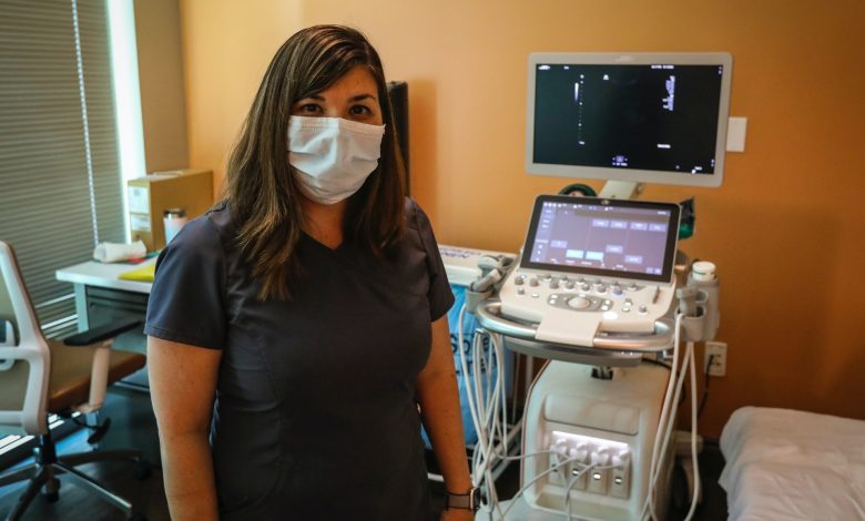
A transducer probe, a central processing unit (CPU), a display, a keyboard with control knobs, disc storage devices, a printer, and other components make up an ultrasound scanner. A sonographer is a person who performs an ultrasound scan, although radiologists, cardiologists, and other experts analyse the findings.
Ultrasound waves are generate by a transducer, which may both emit and detect ultrasound echoes reflect. Ultrasound is a kind of sound that travels through soft tissue and fluids yet returns as echoes from denser things.
This is how a photograph is made. In most cases, ultrasonic transducers’ active components are made of piezo electrics, which are unique ceramic crystal materials. When some materials are expose to an electric field, they can generate sound waves, but they can also generate an electric field when a sound wave impacts them. A gel is squirt into the patient’s stomach.
This prevents air pockets from developing between the transducer and the skin, obstructing the passage of ultrasound waves into the body. If you are looking for ultrasound scans, you may get in touch with the best radiologist in Noida, Dr Ashish Arora, Ultrasound & Imaging Centre.
During The Scan
The sonographer generally holds the transducer, which is softly move over the stomach like a wand. Your ultrasound done vaginally if you are extremely early in your pregnancy, overweight, or have a deep pelvis. A vaginal probe will be place into your vagina to obtain a clear picture of your baby in this case. This will not damage your baby, although it may cause you some discomfort. Ultrasound frequencies for diagnostic purposes are generally between 2 and 18 megahertz (MHz).
These echoes are create when sound waves penetrate through the skin and bounce off fluids, bone, tissue, and organs. The more the ultrasonic bounces back from a dense object, the more it bounces back. The echo, or bouncing back, is what gives the ultrasound picture its characteristics.
Different hues of grey correspond to different densities. The computer’s job is to analyse and transform the waves that return to the probe into electrical signals. The picture is generate using calculations based on the speed of sound and the time it took for the echo to reach the probe. The scanner estimates the distance between the transducer and the tissue border using the speed of sound and the time it takes for each echo to return. Following that, the distances are utilize to create two-dimensional representations of tissues and organs.
Advantages of opting for an Ultrasound Scan
Patients choose ultrasonic scans for a variety of reasons. Examining the developing infant condition of a pregnant woman and determining the due date is quite beneficial. Doctors have also advised patients to undergo an Ultrasound Scan to examine internal organs. Such as the pancreas or liver, or a Doppler ultrasound to monitor blood flow if the patient has a blood circulation problem.
If you are seeking the Best Ultrasound clinic near you in Noida, you may contact Dr Ashish Arora’s clinic, which is outfitt with the most sophisticated technology.
Takeaway
So, do you understand how ultrasonography works now? Although they are safe, screening a pregnant woman unnecessarily. It become necessary if it is medically essential during pregnancy. Anyone who is allergic to latex should tell their doctor so that a latex-covered probe is not used.
For any queries, reach out to the best radiologist in Noida in Sector 19, Dr Ashish Arora clinic.


